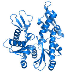
MreB

MreB is a protein found in bacteria that has been identified as a homologue of actin, as indicated by similarities in tertiary structure and conservation of active site peptide sequence. The conservation of protein structure suggests the common ancestry of the cytoskeletal elements formed by actin, found in eukaryotes, and MreB, found in prokaryotes.[1] Indeed, recent studies have found that MreB proteins polymerize to form filaments that are similar to actin microfilaments. It has been shown to form multilayer sheets comprising diagonally interwoven filaments in the presence of ATP or GTP.[2]
Function
MreB controls the width of rod-shaped bacteria, such as Escherichia coli. A mutant E. coli that creates defective MreB proteins will be spherical instead of rod-like. Also, bacteria that are naturally spherical do not have the gene encoding MreB. Prokaryotes carrying the mreB gene can also be helical in shape. MreB has long been thought to form a helical filament underneath the cytoplasmic membrane, however, this model has been brought into question by three recent publications showing that filaments cannot be seen by electron cryotomography and that GFP-MreB can be seen as patches moving around the cell circumference. It has been shown to interact with several proteins that are proven to be involved in length growth (for instance PBP2). Therefore, it probably directs the synthesis and insertion of new peptidoglycan building units into the existing peptidoglycan layer to allow length growth of the bacteria.
References
- ↑ Gunning PW, Ghoshdastider U, Whitaker S, Popp D, Robinson RC (2015). "The evolution of compositionally and functionally distinct actin filaments". J Cell Sci. doi:10.1242/jcs.165563. PMID 25788699.
- ↑ Popp, D; Narita, A; Maeda, K; Fujisawa, T; Ghoshdastider, U; Iwasa, M; Maéda, Y; Robinson, R. C. (2010). "Filament structure, organization, and dynamics in MreB sheets". Journal of Biological Chemistry. 285 (21): 15858–65. doi:10.1074/jbc.M109.095901. PMC 2871453
 . PMID 20223832.
Sources
- Erickson H (2001). "Cytoskeleton. Evolution in bacteria". Nature. 413 (6851): 30. doi:10.1038/35092655. PMID 11544510. - source of information added to this entry as of February 20, 2006
- Swulius; et al. (2011). "Long helical filaments are not seen encircling cells in electron cryotomograms of rod-shaped bacteria.". 407 (4): 650–5. doi:10.1016/j.bbrc.2011.03.062. PMC 3093302
 . PMID 21419100.
 - Dominguez-Escobar; et al. (2011). "Processive movement of MreB-associated cell wall biosynthetic complexes in bacteria.". 333 (6039): 225–8. doi:10.1126/science.1203466. PMID 21636744.
- Garner; et al. (2011). "Coupled, circumferential motions of the cell wall synthesis machinery and MreB filaments in B. subtilis.". 333 (6039): 222–5. doi:10.1126/science.1203285. PMC 3235694
 . PMID 21636745.
