Superficial epigastric artery
| Superficial epigastric artery | |
|---|---|
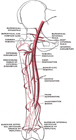
 Scheme of the femoral artery. (Superficial epigastric visible at upper left.) | |
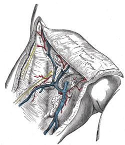
 The left femoral triangle. (Superficial epigastric vesseles labeled at center top.) | |
| Details | |
| Source | Femoral artery |
| Vein | Superficial epigastric vein |
| Identifiers | |
| Latin | Arteria epigastrica superficialis |
| MeSH | A07.231.114.330 |
| TA | A12.2.16.011 |
| FMA | 20734 |
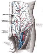
The superficial epigastric artery (not to be confused with the superior epigastric artery) arises from the front of the femoral artery about 1 cm below the inguinal ligament, and, passing through the femoral sheath and the fascia cribrosa, turns upward in front of the inguinal ligament, and ascends between the two layers of the superficial fascia of the abdominal wall nearly as far as the umbilicus.
It distributes branches to the superficial subinguinal lymph glands, the superficial fascia, and the integument; it anastomoses with branches of the inferior epigastric, and with its fellow of the opposite side.
Additional images
 The subcutaneous inguinal ring.
 The femoral artery.
 The great saphenous vein and its tributaries at the fossa ovalis.
 The great saphenous vein and its tributaries.
 The femoral vein and its tributaries.
- Anterior abdominal wall.Intermediate dissection.Anterior view
References
This article incorporates text in the public domain from the 20th edition of Gray's Anatomy (1918)
External links
- Anatomy photo:35:02-0100 at the SUNY Downstate Medical Center - "Anterior Abdominal Wall: Blood Vessels in the Superficial Fascia"
- Anatomy image:7131 at the SUNY Downstate Medical Center
- Anatomy image:7280 at the SUNY Downstate Medical Center
This article is issued from Wikipedia - version of the 4/18/2015. The text is available under the Creative Commons Attribution/Share Alike but additional terms may apply for the media files.