Pupillary response
.jpg)
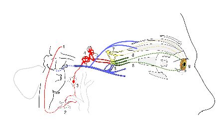
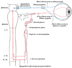
Pupillary response is a physiological response that varies the size of the pupil, via the optic and oculomotor cranial nerve. A constriction response (miosis),[1] is the narrowing the pupil, or which may be caused by scleral buckles or drugs such as opiates/opioids or anti hypertension medications. A dilation response (mydriasis), is the widening the pupil and may be caused by anticholinergic agents or drugs such as MDMA, cocaine and amphetamines. Dilation of the pupil occurs when the smooth cells of the radial muscle, controlled by the sympathetic nervous system (SNS), contract. Constriction of the pupil occurs when the circular muscle, controlled by the parasympathetic nervous system (PSNS), contracts.
The responses can have a variety of causes, from an involuntary reflex reaction to exposure or inexposure to light — in low light conditions a dilated pupil lets more light into the eye — or it may indicate interest in the subject of attention, or sexual stimulation.[2] The pupils contract immediately before REM sleep begins.[3] A pupillary response can be intentionally conditioned as a Pavlovian response to some stimuli.[4]
The latency of pupillary response (the time in which it takes to occur) increases with age.[5] Use of central nervous system stimulant drugs and some hallucinogenic drugs can cause dilation of the pupil.[6]
In ophthalmology, intensive studies of pupillary response are conducted via videopupillometry.[7]
Anisocoria is the condition of one pupil being more dilated than the other.


| Constriction | Dilation | |
|---|---|---|
| Muscular mechanism | Relaxation of iris dilator muscle, activation of iris sphincter muscle | Activation of iris dilator muscle, relaxation of iris sphincter muscle |
| Cause in pupillary light reflex | Increased light | Decreased light |
| Other physiological causes | Accommodation reflex | Fight-or-flight response |
| Corresponding non-physiological state | Miosis | Mydriasis |
See also
References
- ↑ Ellis CJ (November 1981). "The pupillary light reflex in normal subjects" (PDF). Br J Ophthalmol. 65 (11): 754–9. doi:10.1136/bjo.65.11.754. PMC 1039657
 . PMID 7326222.
 - ↑ "Pupil Size as Related to Interest Value of Visual Stimuli", Science, 132 (3423): 349–50, 5 August 1960, doi:10.1126/science.132.3423.349, PMID 14401489
- ↑ "Pupillary Movements During Acute and Chronic Fatigue: A New Test for the Objective Evaluation of Tiredness" (PDF), Investigative Ophthalmology, St. Louis: C.V. Mosby Company, 2 (2): 138–157, April 1963
- ↑ Baker, Lynn Erland (1938). "The Pupillary Response Conditioned to Subliminal Auditory Stimuli". Ohio State University.
- ↑ Latency of pupillary reflex to light stimulation and its relationship to aging (PDF), Federal Aviation Agency, Office of Aviation Medicine, Georgetown Clinical Research Institute, September 1965, p. 12, OCLC 84657376
- ↑ Jaanus SD (1992), "Ocular side effects of selected systemic drugs", Optom Clin, 2 (4): 73–96, PMID 1363080
- ↑ "A new videopupillography", Ophthalmologica, 160 (4): 248–259, 1970, doi:10.1159/000305996, PMID 5439164