Phantom eye syndrome
| Phantom eye syndrome | |
|---|---|
 | |
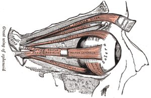
| Anatomy of the eye. The external eye muscles are shown in red. | |
| Classification and external resources | |
| Specialty | neurology |
| ICD-10 | G54.6, G54.7 |
| ICD-9-CM | 353.6 |
The phantom eye syndrome (PES) is a phantom pain in the eye and visual hallucinations after the removal of an eye (enucleation, evisceration).
Symptomatology
Many patients experience one or more phantom phenomena after the removal of the eye:
- Phantom pain in the (removed) eye (prevalence: 26%)[1][2]
- Non-painful phantom sensations[1][2]
- Visual hallucinations. About 30% of patients report visual hallucinations of the removed eye.[1] Most of these hallucinations consist of basic perceptions (shapes, colors). In contrast, visual hallucinations caused by severe visual loss without removal of the eye itself (Charles Bonnet syndrome) are less frequent (prevalence 10%) and often consist of detailed images.
Pathogenesis
Phantom pain and non-painful phantom sensations
Phantom pain and non-painful phantom sensations result from changes in the central nervous system due to denervation of a body part.[3][4] Phantom eye pain is considerably less common than phantom limb pain. The prevalence of phantom pain after limb amputation ranged from 50% to 78%. The prevalence of phantom eye pain, in contrast, is about 30%.
Post-amputation changes in the cortical representation of body parts adjacent to the amputated limb are believed to contribute to the development of phantom pain and non-painful phantom sensations. One reason for the smaller number of patients with phantom eye pain compared with those with phantom limb pain may be the smaller cortical somatosensory representation of the eye compared with the limbs.
In limb amputees, some,[5] but not all, studies have found a correlation between preoperative pain in the affected limb and postoperative phantom pain. There is a significant association between painful and non-painful phantom experiences, preoperative pain in the symptomatic eye and headache.[6] Based on the present data, it is difficult to determine if headaches or preoperative eye pain play a causal role in the development of phantom phenomena or if headache, preoperative eye pain, and postoperative phantom eye experiences are only epiphenomena of an underlying factor. However, a study in humans demonstrated that experimental pain leads to a rapid reorganization of the somatosensory cortex.[7] This study suggests that preoperative and postoperative pain may be an important co-factor for somatosensory reorganization and the development of phantom experiences.
Visual hallucinations
Enucleation of an eye and, similarly, retinal damage, lead to a cascade of events in the cortical areas receiving visual input. Cortical GABAergic (GABA: Gamma-aminobutyric acid) inhibition decreases, and cortical glutamatergic excitation increases, followed by increased visual excitibility or even spontaneous activity in the visual cortex.[8] It is believed that spontaneous activity in the denervated visual cortex is the neural correlate of visual hallucinations.
Treatment
Treatment of painful phantom eye syndrome is provision of ocular prosthesis in the empty orbit.[2]
See also
References
- 1 2 3 Sörös, P.; O. Vo; I.-W. Husstedt; S. Evers & H. Gerding (May 2003). "Phantom eye syndrome: Its prevalence, phenomenology, and putative mechanisms". Neurology. 60 (9): 1542–3. doi:10.1212/01.wnl.0000059547.68899.f5. PMID 12743251. Retrieved 2008-09-23.
- 1 2 3 Shah S.I.A (1994). "Painful Phantom Eye" (PDF). Pk J Ophthalmol. 10 (4 (Index Issue)): 77–78.
- ↑ Ramachandran, Vilayanur S.; W Hirstein (September 1998). "The perception of phantom limbs. The D. O. Hebb lecture". Brain. 121 (9): 1603–30. doi:10.1093/brain/121.9.1603. PMID 9762952. Retrieved 2008-09-23.
- ↑ Nikolajsen, L.; T. S. Jensen (July 2001). "Phantom limb pain". British Journal of Anaesthesia. 87 (1): 107–16. doi:10.1093/bja/87.1.107. PMID 11460799. Retrieved 2008-09-23.
- ↑ Nikolajsen L, Ilkjaer S, Krøner K, Christensen JH, Jensen TS (September 1997). "The influence of preamputation pain on postamputation stump and phantom pain". Pain. 72 (3): 393–405. doi:10.1016/S0304-3959(97)00061-4. PMID 9313280. Retrieved 2008-09-23.
- ↑ Nicolodi, M.; R. Frezzotti; A. Diadori; A. Nuti; F. Sicuteri (June 1997). "Phantom eye: features and prevalence. The predisposing role of headache". Cephalalgia. 17 (4): 501–4. doi:10.1046/j.1468-2982.1997.1704501.x. PMID 9209770. Retrieved 2008-09-23.
- ↑ Sörös, Peter; Stefan Knecht; Carsten Bantel; Tanya Imai; Rainer Wüsten; Christo Pantev; Bernd Lütkenhöner; Hartmut Bürkle; Henning Henningsen (February 2001). "Functional reorganization of the human primary somatosensory cortex after acute pain demonstrated by magnetoencephalography". Neuroscience Letters. 298 (3): 195–8. doi:10.1016/S0304-3940(00)01752-3. PMID 11165440. Retrieved 2008-09-23.
- ↑ Eysel, Ulf T.; Georg Schweigart; Thomas Mittmann; Dirk Eyding; Ying Qu; Frans Vandesande; Guy Orban; Lutgarde Arckens (1999). "Reorganization in the visual cortex after retinal and cortical damage". Restorative Neurology and Neuroscience. 15 (2–3): 153–64. PMID 12671230. Retrieved 2008-09-23.
External links
- Cole, Jonathan. "Phantom limb pain". Wellcome Trust. Retrieved 2008-09-23.